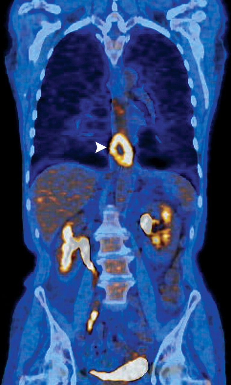
The basic principle behind a ct scan relies on the reconstruction of a three dimensional image of an organ, by a computer. Pet scan is the shorten form of positron emission tomography.
Pet scans use a radioactive tracer to show how an organ is functioning in real time.

Is cat scan and pet scan the same. It is only with a pet/ct scan that you will know for sure. Why pet scans are used. An effective dose of about 7 msv for a whole body study).
Usually fdg (fluro deoxy glucose) is used. Â this type of examination helps to evaluate any damage that presents in the bones. Combined with an mri scan, a pet scan can produce multidimensional images showing the inside of the human body.
This tracer travels through your body systems. What is a ct scan? Ct scan was initially known as the emi scan because of the company name that introduced this equipment in the market.
A pet scan, on the other hand, shows doctors how the tissues in your body work on a cellular level. The pet scan uses a mildly radioactive drug to show up areas of your body where cells are more active than normal. The main difference in a pet scan vs.
Physicians order each type of scan for different purposes. This combination test produces 3d images that allow for a more accurate diagnosis. Scan is a process that examines the body’s internal bone structure using nuclear scanning.
You’ll receive a substance called a “tracer” containing glucose with a little bit of radioactive material attached before your test. This will emit the positrons. The beauty of the axumin pet scan is that it offers the possibility of detecting small metastatic lesions in the lymph nodes with psa levels in the 1 to 10 range.
While they can be ordered in combination with one another, pet scans and ct scans do have distinct diagnostic purposes. One actually cost more and takes more time to do than the other. The pet scan works by using radiation to show activity within the body on a cellular level.
While ct and mri scans show images of your body’s internal organs and tissues, pet scans can give your healthcare provider a view of complex systemic diseases by showing problems at the cellular. Perhaps the main difference between a ct scan and a pet scan is their focus. Ct scan was initially generally called the emi scan because of the company title that launched this gear on the market.
The same assessment applies to tracking the. It acts like a dye for the imaging scan to pick up on. A radiographer operates the scanner.
Pet scan images can detect cellular changes in organs and tissues earlier than ct and mri scans. Some of the reasons for ordering a pet scan, according to johns hopkins, include: The pet and ct scans are done at the same time on the same machine.
A positron emission tomography (or pet scan) is mainly used in cancer treatment, neurology, and cardiology. What is the difference between a ct and pet scan? A pet scan can show how well certain parts of your body are working, rather than simply showing what they look like.
Pet/ct scans provide significantly more information than ct scans, and are far more reliable when diagnosing cancer. On the other hand, a pet scan takes anywhere between 2 to 4 hours. Â a pet scan, also known as positron emission topography, does not use nuclear.
Pet scans are often combined with ct scans to produce even more detailed images. Historically, simple bone scans and cat scans required psa levels in the 10 to 50 range before enough cancer would be present to be detected on a scan. The isotopes (the atoms can divide and emit the rays) used in the pet scan.
This sugar substance is taken up by cells that use the most energy. The level of tumor metabolism is compared on pet scans taken before and after chemotherapy. When compared directly, the ct scan and pet scan look similar because they both involve the use of contrasting agents, but the type you get depends on your scan.
Pet scans, ct (computerized tomography), and mris are similar in many ways. A small amount of a radioactive sugar substance is injected into the patient’s body. Bone scan vs pet scan a bone.
The pet ct scan helps the physician to see the level of activity of certain body organs and tissues, along with their structure. Â this allows the identification of bone growth or perhaps the breakdown of these in the human body. Using nuclear medicine these exams allow particular focus on oncological symptoms in the brain and heart as well as any vascular or tissue abnormalities.
Positron is like an electron in weight but positively charged. The radiation dose from such a scan can be low (e.g. The physician is able to precisely overlay the metabolic data of the pet scan and the detailed anatomic data of the ct scan to make a more detailed image than either test would make by itself.
Pet scans are excellent at analyzing the biological processes of the body and at detecting pathology such as cancer at the very earliest stages. Pet scans are typically used in conjunction with a ct scan or mri. Because of the axial image resolutions, the term also gets known as cat scan.
The difference between and cat scan and a pet scan is that a cat scans the outline of the bones in the body and a pet scans the biological processes of the body. The reality is that you cannot rely on a ct scan (or ultrasound, mri, or blood test) to tell you if you have cancer. Positron is emitted during nuclear reactions.
A successful response seen on a pet scan frequently precedes alterations in anatomy and would therefore be an earlier indicator of tumor shrinkage than would be seen with other diagnostic modalities.
 PET/CT Basics Pet ct, Pet scan, Pets
 Coronal image of a CT abdomen and pelvis with IV contrast
 Idea by Shon Duplechian on pet scan Pet scan, Pet ct
 Chest CT scan in a patient with cough shows a
 Abdomen CT scan in a child shows an abnormal kidney
 PET/CT Diagnostic Imaging Services Pet ct, Diagnostic
 CT examination of the neck are done with the IV
 Chest CT Scan Imaging RadTechOnDuty Radiology, Anatomy
 PET CT Scan in Delhi 9,999 Ct scan, Pet ct, Pet scan
 rabbit undergoing CT scan at Anton Vets in UK Pet rabbit
 CT scan of the same patient as in Fig.15 showing the
 XRay, MRA, MRI, PET SCAN and CT images are all different
 Multidetector CT of the chest shows multiple cavitary
 Chest CT Scan Imaging RadTechOnDuty Ct scan
 What are the structures labelled AF in this axial


